
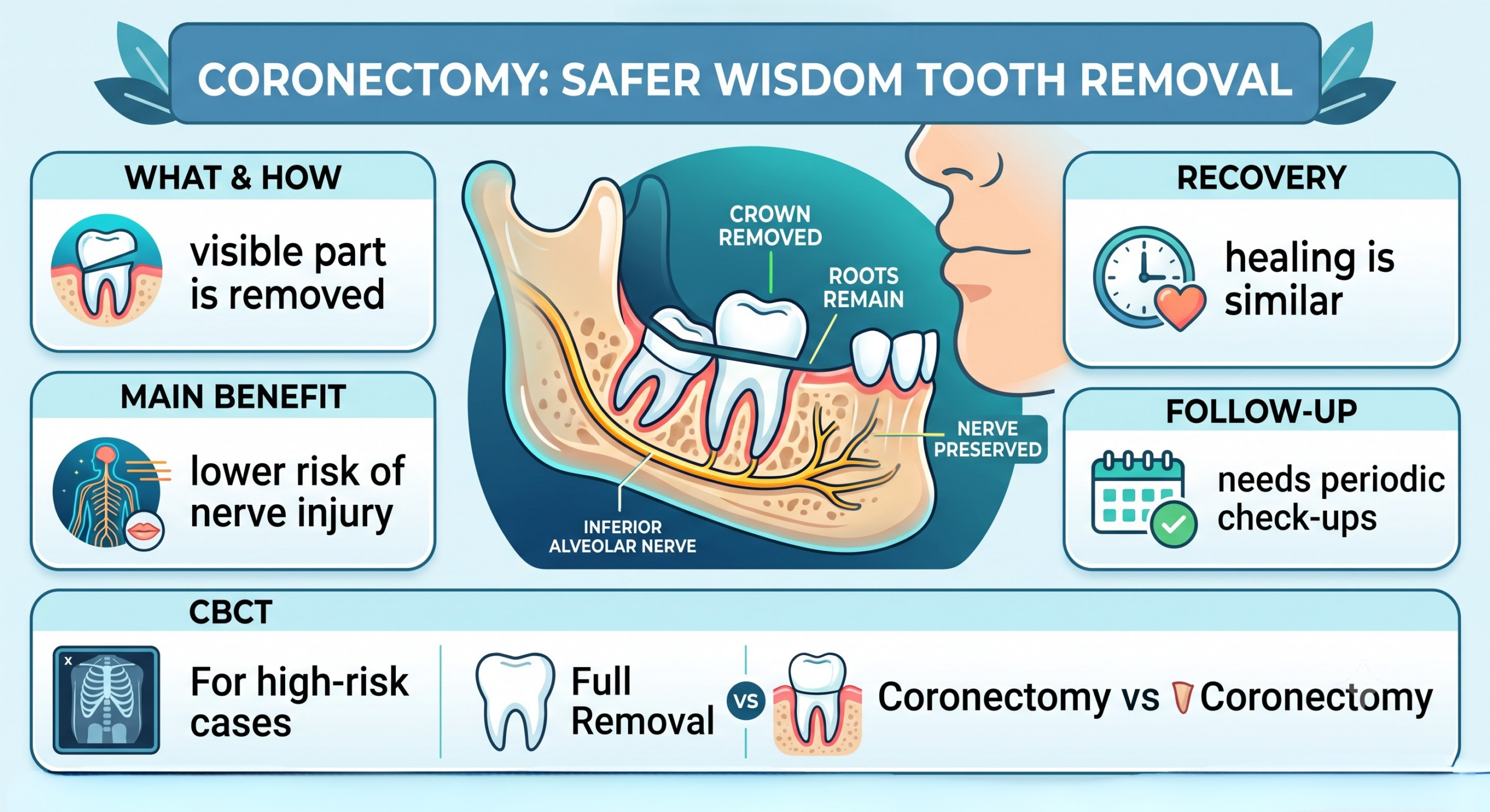
A coronectomy is a wisdom tooth procedure that removes the crown of an impacted tooth while deliberately leaving the roots in place, and it’s changing how dentists handle high-risk extractions near the nerve.
If your dentist has ever hesitated before pulling a wisdom tooth, mentioning something about “nerve proximity” or “risk of numbness,” you’ve likely brushed up against the exact scenario coronectomy was designed to solve. It isn’t a shortcut or a compromise. It’s a deliberate, evidence-backed technique that oral and maxillofacial surgeons reach for when a full extraction of a mandibular third molar could do more harm than good.
This guide walks through what coronectomy actually involves, who it’s meant for, how it compares to standard extraction, and what recovery looks like so you can walk into your consultation with clear, informed questions.
What Is a Coronectomy?
A coronectomy, sometimes called an intentional partial odontectomy, is a surgical technique where the dentist removes only the crown (the visible part) of an impacted mandibular third molar. The roots stay embedded in the jawbone rather than being extracted.
This isn’t laziness or an incomplete job. It’s a purposeful decision made when the roots of the wisdom tooth sit dangerously close to the inferior alveolar nerve, a branch of the trigeminal nerve responsible for sensation in your lower lip, chin, and part of your tongue. Pulling those roots out could stretch, compress, or sever that nerve, leading to paresthesia, the clinical term for numbness or altered sensation that can be temporary or, in rare cases, permanent. The lingual nerve, which runs close to the surgical site, is also at risk during standard extraction, making careful technique essential regardless of which approach is used.
By leaving the roots undisturbed, the dentist sidesteps that risk entirely while still eliminating the crown, which is usually what’s causing pain, infection, or crowding in the first place.
Coronectomy vs. Full Wisdom Tooth Extraction
How the Procedures Differ
A standard wisdom tooth extraction removes the entire tooth, crown and root, from the socket. It’s the go-to approach for most impacted teeth because it’s definitive: once the tooth is out, there’s nothing left to monitor or worry about down the line.
A coronectomy, on the other hand, only takes the crown. The roots are trimmed slightly below the bone level and left to remain, often permanently, though in some cases they migrate away from the nerve over time through natural bone remodeling and can be removed later with far less risk.
| Feature | Full Extraction | Coronectomy |
| What’s removed | Entire tooth (crown + roots) | Crown only |
| Nerve risk | Higher when roots are near the inferior alveolar nerve | Significantly lower |
| Follow-up needed | Rarely | Sometimes, to monitor root migration via imaging |
| Healing time | 1-2 weeks typical | Similar, sometimes slightly longer |
| Best for | Low-risk impactions | High-risk, nerve-adjacent impactions |
Why Dentists Choose Coronectomy for High-Risk Cases
Oral and maxillofacial surgeons don’t reach for coronectomy as a first option. It’s specifically reserved for situations where imaging shows the roots overlapping or wrapping around the inferior alveolar nerve canal. In those cases, research has consistently shown that coronectomy carries a dramatically lower rate of paresthesia compared to complete extraction, while still resolving the pain and infection risk tied to the crown.
Who Needs a Coronectomy?
Wisdom Teeth Close to the Inferior Alveolar Nerve
Not every impacted wisdom tooth qualifies for this approach. Coronectomy is typically recommended when a lower wisdom tooth’s roots are intimately associated with the nerve canal, meaning there’s little to no safe margin for a surgeon to work around during a full extraction. Impacted teeth are generally classified by their angulation, mesioangular, distoangular, vertical, or horizontal, and this classification helps surgeons predict both surgical difficulty and nerve proximity.
Younger patients, whose roots haven’t fully matured, are sometimes better candidates because incomplete root formation can make full extraction safer. Still, for many adults with deeply impacted lower third molars, coronectomy remains the safer path.
Signs on Imaging That Point to Coronectomy
Dentists rely on imaging, often a panoramic radiograph (OPG) followed by cone-beam computed tomography (CBCT), to assess the tooth’s relationship to the nerve. CBCT scans, in particular, reveal the three-dimensional relationship between the root and the nerve canal far more precisely than a flat panoramic image. Certain findings raise red flags for extraction risk:
- Darkening or interruption of the nerve canal’s white outline where it crosses the roots
- Narrowing of the nerve canal itself
- Deflection or curving of the root around the canal
- Direct root grooving from the canal
When two or more of these signs appear together, a surgeon is far more likely to recommend coronectomy over standard extraction.
The Coronectomy Procedure Step by Step
Before the Procedure
Your dentist or oral and maxillofacial surgeon will typically order a CBCT scan to map the exact position of the roots relative to the nerve. You’ll discuss anesthesia options, which can range from local anesthesia (commonly lidocaine) to sedation or general anesthesia depending on the complexity of the case and your comfort level. Expect a conversation about medical history, current medications, and any allergies.
Coronectomy isn’t appropriate for every patient, even those who are otherwise nerve-risk candidates. It’s generally contraindicated when the tooth roots are mobile, when there’s active infection or pericoronitis at the surgical site, or when the tooth is horizontally impacted with roots positioned in a way that crown removal could still disturb them. Your surgeon will screen for these factors before recommending the procedure.
During the Procedure
The surgeon numbs or sedates you, then makes an incision to access the tooth. Rather than working the entire tooth loose, they section the crown away from the roots using a surgical drill, remove the crown, and trim the remaining root surface down to about 3-4 millimeters below the bone crest. The area is then cleaned, and the gum tissue is sutured closed over the retained roots.
The whole procedure usually takes a similar amount of time to a standard surgical extraction, often somewhere between 20 and 60 minutes depending on the tooth’s position and complexity.
After the Procedure
You’ll go home with the same general aftercare instructions as any oral surgery: ice packs to control swelling, a soft food diet for the first few days, chlorhexidine mouthwash to keep the surgical site clean, and pain relief such as ibuprofen or acetaminophen as needed. Your dentist may also prescribe antibiotics if there’s any concern about infection risk. Your surgeon will schedule follow-up appointments, sometimes over months or years, to keep an eye on the retained roots via periodic CBCT or panoramic imaging.
Benefits of Coronectomy
Coronectomy offers several advantages that make it worth considering when you’re a candidate:
- Dramatically reduced paresthesia risk. This is the headline benefit and the entire reason the procedure exists.
- Resolves pain and infection. Removing the crown eliminates the source of most day-to-day symptoms, including pericoronitis, the gum inflammation that often develops around a partially erupted wisdom tooth.
- Comparable recovery time. Healing generally mirrors a standard extraction.
- Root migration over time. In many patients, the retained roots gradually move away from the nerve through natural bone remodeling, sometimes allowing for safer removal later if needed.
- Lower long-term complication rate for patients who were genuinely high-risk candidates for full extraction.
Risks and Limitations of Coronectomy
No procedure is without trade-offs, and coronectomy has a few worth understanding upfront.
Root Migration
While root migration away from the nerve is common and considered a positive outcome, it doesn’t happen predictably in every patient. Some roots stay put, and a small percentage may migrate toward the surface instead, which can occasionally cause them to become exposed, infected, or a source of localized pericoronitis.
Need for a Second Surgery
In a minority of cases, retained roots become symptomatic later on, whether due to infection, migration toward the gum surface, or patient discomfort. When that happens, a second, usually much safer, procedure may be needed to remove the roots once they’ve moved away from the nerve.
Coronectomy Recovery Timeline
Most patients follow a fairly predictable healing curve:
- Days 1-3: Swelling and mild-to-moderate discomfort peak, managed with ice and pain medication.
- Days 4-7: Swelling subsides noticeably, and most people return to normal eating and speaking. This is also the window when dry socket, or alveolar osteitis, would typically appear if it’s going to occur, though it’s less common with coronectomy since the socket isn’t left empty the way it is after full extraction.
- Week 2: Sutures are typically checked or dissolve on their own, depending on the type used.
- Months 3-6: First follow-up CBCT or panoramic imaging to check root position and healing.
- Ongoing: Periodic imaging, often annually, to track any root migration.
Most people find the recovery experience nearly identical to a standard wisdom tooth extraction, aside from the longer-term monitoring schedule.
Coronectomy vs. Leaving the Tooth Alone
It’s fair to ask whether doing nothing is an option. In some cases, if the wisdom tooth is asymptomatic and not causing crowding, decay, or infection, a dentist may recommend simple observation. However, if the tooth is actively causing pain, cavities, gum infection, pericoronitis, or damage to neighboring teeth, leaving it untreated isn’t a safe long-term strategy. Coronectomy offers a middle path: it addresses the problematic crown without taking on the nerve risk of full extraction.
Cost of Coronectomy
Coronectomy costs vary widely depending on your location, the complexity of the case, and whether you need sedation or general anesthesia. In general, it falls in a similar price range to a surgical wisdom tooth extraction, though the additional CBCT imaging and follow-up visits can add to the overall cost over time. It’s worth checking with your dental insurance provider, since coverage for coronectomy can differ from standard extraction coverage. The American Association of Oral and Maxillofacial Surgeons offers general guidance on wisdom tooth procedure costs and considerations that’s worth reviewing before your consultation, and the American Dental Association is another trusted resource for understanding typical procedure standards.
Frequently Asked Questions
1. Is a coronectomy painful?
Most patients report discomfort similar to a standard extraction, well managed with prescribed or over-the-counter pain relief like ibuprofen or acetaminophen during the first few days.
2. Will the leftover roots cause problems later?
In most cases, no. The roots typically remain stable or migrate safely away from the nerve. A small percentage of patients need a follow-up procedure if the roots become symptomatic or develop pericoronitis.
3. How do I know if I’m a candidate for coronectomy?
Your oral and maxillofacial surgeon will determine this through imaging, usually a CBCT scan, to assess how close your tooth’s roots are to the inferior alveolar nerve and to rule out contraindications like active infection or root mobility.
4. Does insurance cover coronectomy?
Coverage varies by provider and plan. It’s best to confirm directly with your insurance company before scheduling.
5. How long does the coronectomy procedure take?
Most procedures take between 20 and 60 minutes, depending on the tooth’s position and the complexity of the case.
6. Can coronectomy be converted to a full extraction if something goes wrong?
Yes, in some cases the surgeon may determine mid-procedure that full extraction is safer, particularly if the roots turn out to be mobile once exposed, and this possibility is usually discussed beforehand.
7. Is coronectomy a new procedure?
Coronectomy has been documented in dental literature since the 1980s and has become increasingly common as CBCT imaging technology has improved nerve-risk assessment.
Conclusion
Coronectomy exists for one clear reason: to protect the inferior alveolar nerve responsible for feeling in your lip, chin, and tongue when a wisdom tooth’s roots sit too close for a safe full extraction. It isn’t a lesser procedure. It’s a deliberate, well-researched alternative that trades a small, well-managed long-term monitoring commitment for a much lower risk of permanent paresthesia. If your dentist has raised coronectomy as an option, it’s worth taking seriously and worth asking detailed questions about your specific CBCT results and whether any contraindications apply before deciding on your next step.