
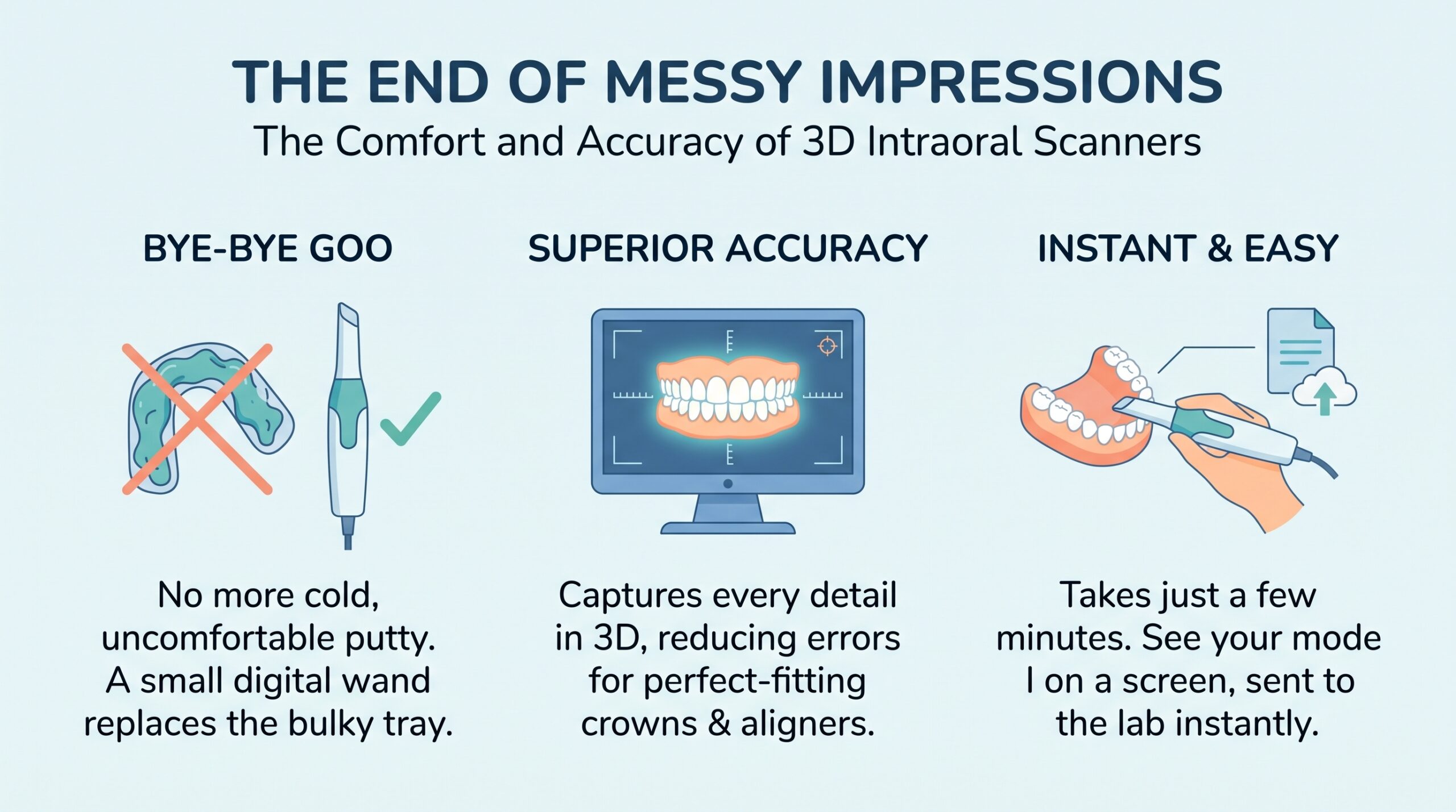
Remember the last time you bit down into a tray of cold, gooey impression material? Most people don’t have fond memories. The gagging, the awkward wait, and the unpleasant taste make it one of dentistry’s least-loved moments. Thankfully, there’s a better way. An intraoral scanner replaces that sticky tray with a small, wand-shaped camera. It captures your teeth in a few minutes and builds a detailed 3D model on a screen.
But here’s the big question: is a digital scan really as accurate as a traditional impression? In many everyday cases, the answer is a confident yes. A modern intraoral scanner can record fine detail, reduce human error, and give dentists a model they can trust. In this guide, we’ll explain how the technology works, why it’s so precise, and what to look for when choosing one. We’ll also answer the questions patients and practice owners ask most.
Whether you’re a dentist weighing an upgrade or a patient curious about your next visit, you’re in the right place. Let’s dive in.
What Is an Intraoral Scanner?
An intraoral scanner is a handheld digital device that captures the shape of your teeth, gums, and nearby soft tissue. Instead of taking a physical mold, it records thousands of tiny images and joins them into one 3D model. Dentists call this process “digital impressioning.”
The technology has come a long way. Early systems were bulky, slow, and very expensive. Today’s devices are lightweight, fast, and far more affordable. As a result, they’ve moved from a “nice to have” gadget to a standard tool in many modern clinics.
Think of it like taking a panoramic photo with your phone. You sweep the camera across a scene, and the software stitches the pieces together. An intraoral scanner does the same thing, only in three dimensions and at a much finer scale.
How an Intraoral Scanner Works
Every intraoral scanner follows the same basic idea: light goes in, data comes out. The wand projects light onto the tooth surface. Tiny sensors then record how that light bounces back. Software turns those readings into millions of data points, which form a “point cloud.” Finally, the program connects those points into a smooth, detailed 3D surface.
Different brands use different methods to capture that light. Here are the most common ones:
- Confocal imaging: Uses focused light at different depths to build a layered picture of the tooth.
- Triangulation: Projects a pattern onto the tooth and measures how the pattern bends.
- Active wavefront sampling: Tracks how light waves move to calculate depth.
- Structured light: Uses patterned light and multiple cameras to capture shape quickly.
Each method has its own strengths. However, they all aim for the same goal, which is a clean, accurate digital copy of your mouth.
During a scan, the dentist moves the wand slowly along your teeth. You’ll usually see the model build up live on a monitor. If the scanner misses a spot, the software shows a gap, so the dentist can go back and fill it in right away. That instant feedback is a huge advantage over a traditional impression, where you often don’t find a flaw until the lab calls.
Key Parts of a Digital Scanning System
An intraoral scanner isn’t just a wand. It’s a small system of parts that work as a team. Knowing these parts helps you understand why results are so reliable.
| Component | What It Does |
| Handheld wand | Captures images of teeth and gums |
| Scanning tip | Sits inside the mouth; often heated or autoclavable to prevent fogging and spread of germs |
| Processing unit | Turns raw images into a 3D model |
| Software | Stitches, cleans, and analyzes the scan |
| Display screen | Shows the live model to the dentist and patient |
| Export file (such as STL or PLY) | Sends the scan to a lab or milling machine |
The software deserves a special mention. Modern programs use smart algorithms to remove extra data, like a cheek or tongue that drifted into view. They also highlight problem areas. In short, the software acts like a second set of eyes.
Once the scan is done, the file travels over the internet to a dental lab in seconds. There’s no box to ship and no waiting for the courier. That alone saves days in many cases.
Why Traditional Impressions Fall Short
To appreciate the accuracy of an intraoral scanner, it helps to look at what it replaces. Traditional impressions have served dentistry well for over a century. Still, they have real limits, and many of those limits come from the materials themselves.
Impression materials like alginate and silicone are soft and flexible. They can stretch, shrink, or warp. They’re also sensitive to temperature, moisture, and timing. Even a skilled dentist can’t control every one of those factors every time.
Common Impression Errors
A traditional impression passes through many hands before it becomes a crown or a retainer. Each step is a chance for something to go wrong. Here are the most common trouble spots:
- Air bubbles: Tiny voids can hide important edges, such as the margin of a prepared tooth.
- Distortion: If the tray moves or the material is pulled out too fast, the mold can warp.
- Material shrinkage: Some materials change shape slightly as they set or while they wait to be poured.
- Saliva and blood: Moisture can blur fine detail and weaken the final cast.
- Stone model errors: The plaster or stone poured into the mold can add its own small mistakes.
- Shipping delays: Time and temperature changes during transport can affect the impression.
When any of these problems appear, the lab might send the case back. The patient then returns for a second try, which costs time and money for everyone. Studies and clinical experience both show that remakes are frustrating for patients and staff alike.
An intraoral scanner avoids many of these issues. There’s no material to shrink, no stone to pour, and no box to ship. The scan is a digital file, so it doesn’t change once it’s saved.
Patient Comfort Problems
Accuracy isn’t the only thing at stake. Comfort matters too, and it affects the result. A patient who’s gagging or tense is more likely to move, and movement ruins a good impression.
Many people have a strong gag reflex. Others feel anxious in the dental chair. For these patients, a mouth full of putty can feel overwhelming. Some even avoid dental visits because of it.
The wand of an intraoral scanner is small and smooth. It doesn’t fill the whole mouth, and there’s no taste or smell. Most patients tolerate it very well. Children, older adults, and anxious patients often find scanning a pleasant surprise. Happier patients stay still, and stillness leads to better data.
The Accuracy of a 3D Intraoral Scanner
Now let’s get to the heart of the matter. How accurate is a 3D scan, really? The short answer: very accurate, especially for the small areas dentists work on every day. The longer answer needs a few definitions, so let’s break it down.
Trueness and Precision Explained
Researchers use two main words to describe accuracy. They’re easy to mix up, so here’s a simple way to remember them.
- Trueness tells you how close a scan is to the real object. Think of hitting the bullseye on a dartboard.
- Precision tells you how consistent repeated scans are. Think of landing all your darts in the same tight spot, even if it’s not the bullseye.
You need both to get a reliable result. A good intraoral scanner is true and precise. It captures the real shape of the teeth, and it does so again and again.
Accuracy is measured in microns. A micron is one-thousandth of a millimeter, which is far thinner than a human hair. Published studies on modern scanners commonly report errors in the range of tens of microns for single teeth and short spans. That’s well within what’s needed to make a crown that fits comfortably.
Of course, results vary by brand, software version, and technique. That’s why it pays to look at independent research rather than rely only on marketing claims.
Single Crowns vs. Full-Arch Scans
Here’s where a little honesty helps. Scan accuracy isn’t identical in every situation. It depends a lot on how much of the mouth you’re capturing.
For a single crown, an inlay, or a short bridge, an intraoral scanner performs extremely well. The area is small, so there’s little room for error to build up. Many studies show results that match or beat traditional impressions in these cases.
For a full arch, the job is harder. The software must stitch together a long, curved shape. Tiny errors can add up as the scan moves from one side to the other. This is called “cumulative error.” Smooth, flat areas, like a toothless ridge, are especially tricky because there are few landmarks to guide the stitching.
The good news is that technology keeps improving. Newer scanners use better cameras and smarter software to lower cumulative error. Many dentists also use special techniques, such as scan paths and reference markers, to boost full-arch results. So while full-arch work needs extra care, it’s very doable with the right tools and training.
What Affects Scan Accuracy
Even the best intraoral scanner can’t work miracles on its own. Several factors shape the final result. Knowing them helps both dentists and patients feel confident.
| Factor | Why It Matters | How to Improve It |
| Operator skill | Scan path and speed affect stitching. | Practice and formal training |
| Moisture | Saliva and blood scatter light. | Dry the area and retract the tissue. |
| Scanner calibration | Drift can reduce accuracy. | Calibrate on schedule. |
| Tooth surface | Shiny or translucent surfaces can confuse sensors. | Use powder or choose a powder-free model. |
| Software version | Updates often boost precision. | Keep software current |
| Scan strategy | A smart path reduces cumulative error. | Follow the maker’s recommended path. |
Notice how many of these are under the dentist’s control. That’s encouraging. With good habits, a team can get consistent, high-quality scans day after day.
Benefits Beyond Accuracy
Accuracy gets the headlines, but it’s only part of the story. An intraoral scanner changes how a whole practice runs. The benefits ripple out to patients, staff, and labs.
Faster Workflow and Fewer Remakes
Time is money in any clinic. A digital scan speeds up almost every step. The dentist can check the scan on the spot and rescan any weak area before the patient leaves. That means fewer surprise callbacks.
The file also reaches the lab almost instantly. There’s no waiting for a courier or for stone to set. Some practices pair their scanner with an in-office milling unit. In that case, a patient can walk in with a damaged tooth and leave with a new crown in a single visit.
Here’s a quick comparison of the two workflows:
| Step | Traditional Impression | Intraoral Scanner |
| Capture | Tray, material, and wait time | Wand sweep in minutes |
| Quality check | Often at the lab | Instant on screen |
| Transfer | Shipped by courier | Sent online in seconds |
| Model | Poured stone cast | Digital file (print if needed) |
| Remake risk | Higher, due to many steps | Lower, with live feedback |
Fewer remakes also mean less waste. There’s less impression material, less packaging, and fewer shipping trips. That’s a small win for the planet, too.
Better Patient Communication
Here’s a benefit many people don’t expect. A digital scan is a powerful teaching tool. When the model appears on a big screen, patients can see their own teeth in 3D. The dentist can point out a cracked tooth, worn enamel, or a gap that needs attention.
Seeing is believing. Patients who understand their condition are more likely to say yes to treatment. They also feel more involved in their own care, which builds trust.
Many systems go a step further. They let the dentist show a simulated “after” smile. For example, an orthodontic patient can preview how their teeth might move over time. That kind of preview turns an abstract plan into something real and exciting.
Orthodontics and Implants
The reach of the intraoral scanner goes far beyond crowns. It’s now central to several fields of dentistry.
Clear aligners. Most clear aligner companies accept digital scans directly. The scan guides the design of each tray in the treatment plan. Because the data is so precise, aligners tend to fit well from the very first set.
Dental implants. Scanners help plan where an implant should sit. When combined with a 3D X-ray, the data lets dentists design surgical guides that improve placement. Special scan bodies capture the exact position of an implant already in the jaw, so the final crown fits properly.
Dentures and sleep devices. Digital scans are also used for dentures, night guards, and sleep apnea appliances. Patients with a strong gag reflex especially appreciate skipping the messy tray.
Restorative care. Inlays, onlays, veneers, and bridges all benefit from a clean digital margin. A sharp, clear edge helps the lab design a restoration that seals tightly against the tooth.
As you can see, one intraoral scanner can support many types of treatment. That versatility is a big reason why so many practices are making the switch.
Choosing the Right Intraoral Scanner
If you’re a dentist or practice manager, you’re probably wondering how to pick a scanner. The market is crowded, and the options can feel overwhelming. Don’t worry. A simple checklist can help you sort things out.
Features to Compare
Start by thinking about how you’ll use the device every day. Then compare models on the points that matter most to your team.
- Accuracy data: Look for independent studies, not just brand claims.
- Scan speed: Faster scanning keeps patients comfortable and saves chair time.
- Wand size and weight: A lighter, slimmer wand reduces hand fatigue and fits small mouths.
- Color scanning: True-color capture helps with shade matching and gum health checks.
- Powder-free option: Skipping the powder saves time and mess.
- Open vs. closed system: Open systems export standard files, so you can work with any lab. Closed systems limit you to one ecosystem.
- Software and updates: Regular updates keep your scanner sharp over time.
- Training and support: Good onboarding makes a big difference in daily results.
- Integration: Check that it plays well with your practice software, milling unit, and 3D printer.
Whenever possible, ask for a live demo. Better yet, try scanning a few real cases. Your own hands and your own patients will tell you more than any brochure.
Cost and Return on Investment
Let’s talk money. An intraoral scanner is a real investment, and prices vary widely by brand and package. Some clinics buy outright, while others lease or pay through a subscription. Be sure to ask about software fees, warranty, and update costs, since these can add up.
The good news is that a scanner can pay for itself. Think about what you save and what you gain:
- Less spending on impression materials and trays
- Lower shipping costs
- Fewer remakes and wasted chair time
- Higher case acceptance from clearer patient communication
- New services, such as aligners and same-day crowns
Every practice is different, so run the numbers for your own situation. Consider how many impressions you take each month and how much each remake costs you. Many dentists find that the savings and new revenue add up faster than they expected.
Training matters here, too. A scanner only delivers value if the team uses it well. Plan time for practice, and don’t be shy about asking the manufacturer for help. A little effort at the start leads to smoother scans and happier patients later.
Frequently Asked Questions
1. Is an intraoral scanner as accurate as a traditional impression?
For most everyday work, yes. Many studies show that an intraoral scanner matches or beats traditional impressions for single crowns and short bridges. Full-arch scans are harder, but modern devices and good technique keep improving the results.
2. Does scanning hurt?
No. The wand is smooth and doesn’t press into your gums. You may feel the tip touching your teeth, and you’ll need to keep your mouth open for a few minutes. Most patients say it’s far more comfortable than a tray of impression material.
3. How long does a digital scan take?
A full-mouth scan often takes only a few minutes. A single tooth can take less than a minute. The exact time depends on the scanner, the case, and the dentist’s experience.
4. Can an intraoral scanner be used for dental implants?
Yes. Dentists use scanners to plan implant placement and to capture the position of an implant already in the jaw. Combined with 3D X-ray data, the scan helps create guides and restorations that fit precisely.
5. Is an intraoral scanner safe?
Yes. These devices use harmless visible light, not X-rays, so there’s no radiation. Clinics also clean or replace the scanning tips between patients to prevent the spread of germs.
6. How much does an intraoral scanner cost?
Prices range widely based on the brand, features, and software plan. Some practices buy outright, while others lease or subscribe. It’s smart to compare total costs, including updates and support, and to estimate your expected savings.
7. Do I need a special lab to use digital scans?
Not usually. Most modern labs accept digital files. If you choose an open-system scanner, you can send files to almost any lab. It’s a good idea to confirm file compatibility before you buy.
Conclusion
The days of gooey trays and gag-inducing impressions are fading fast. An intraoral scanner offers a cleaner, quicker, and more comfortable way to capture your smile. Just as important, it delivers impressive accuracy, especially for the crowns, aligners, and implants that dentists place every day.