
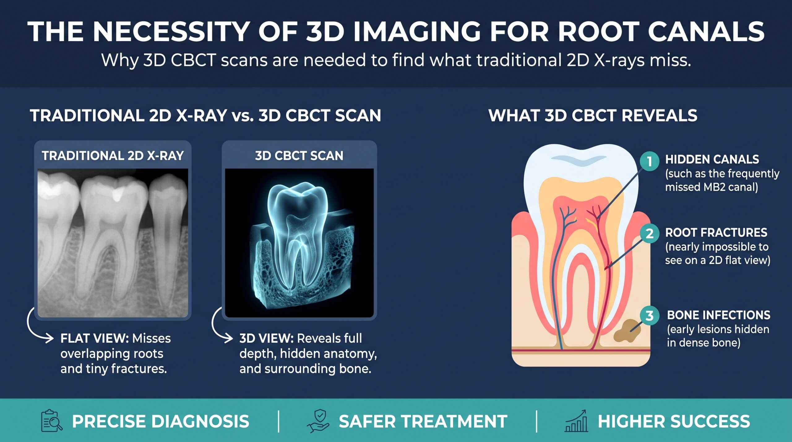
Root canal treatment has a high success rate when every canal is found, cleaned, and sealed. It fails most often for one simple reason: something was left behind. A canal that was never located, a fracture nobody saw, or an infection that spread farther than the X-ray suggested can all quietly undermine an otherwise well-performed procedure.
This is where CBCT in endodontics changes the picture. Cone beam computed tomography gives your endodontist a three-dimensional view of a tooth, its roots, and the surrounding bone. It reveals hidden root canal anatomy that a standard dental X-ray simply cannot show.
This article explains what CBCT is, why 2D X-rays fall short, which complex cases benefit most, and what to expect if your dentist recommends a scan.
What Is CBCT, and How Does It Work?
CBCT stands for cone beam computed tomography. The machine rotates around your head once, capturing a cone-shaped beam of low-dose X-rays from many angles. Software then reconstructs those images into a 3D volume.
The endodontist can scroll through that volume slice by slice, in three planes:
- Axial (a horizontal cross section, like looking down the length of the root)
- Coronal (a front-to-back view)
- Sagittal (a side-to-side view)
This lets the clinician examine one tooth from every direction without any overlapping structures getting in the way.
How CBCT Differs From a Medical CT Scan
A medical CT scanner is built for soft tissue and uses a fan-shaped beam. CBCT is designed for hard tissue such as teeth and bone. It generally delivers a lower radiation dose, uses a smaller and more affordable machine, and produces very high resolution images of tiny structures like root canals and the periodontal ligament space.
Limited Field of View Scans
For endodontic use, the scan is usually limited to a small field of view. That means only the tooth or teeth of interest are imaged, not the entire jaw. A smaller field means a smaller radiation dose and sharper detail where it matters.
Why Traditional 2D X-Rays Miss So Much
Periapical and bitewing radiographs remain essential tools. They are fast, inexpensive, and excellent for many diagnoses. But they have a built-in limitation: they compress a three-dimensional object into a flat image.
Superimposition and Anatomical Noise
When X-rays pass through a tooth, the roots, surrounding bone, the zygomatic arch, and the sinus floor all stack on top of each other in the final image. This is called anatomical noise. It can hide a canal behind another canal, mask the edge of an infection, or make a healthy area look diseased.
Geometric Distortion
A 2D image can also stretch, shorten, or misrepresent the true length and angle of a root. Small changes in the angle of the film or sensor can change how the anatomy appears.
Small Lesions Hide in Dense Bone
Research comparing 2D and 3D imaging has repeatedly shown that CBCT detects periapical lesions that conventional radiographs miss, especially when the infection sits in or near thick cortical bone. A tooth can look normal on a standard film while a lesion is already developing around the root tip.
Hidden Root Canal Anatomy CBCT Helps Reveal
Teeth are not simple tubes. Canals branch, curve, split, and rejoin. Many of the most common reasons for retreatment come from variations that are almost impossible to confirm on a flat image.
The Second Mesiobuccal Canal (MB2)
The upper first molar is the classic example. Its mesiobuccal root very often contains a second canal, called MB2. Studies using CBCT and magnification report it in a large majority of these teeth. It is also one of the most frequently missed canals in endodontic failure. A missed MB2 leaves infected tissue inside the tooth, and symptoms or a persistent lesion often follow.
Middle Mesial Canals in Lower Molars
Lower first molars can have an extra canal between the usual mesiobuccal and mesiolingual canals. It is a small anatomical detail, but it can harbor bacteria if untreated.
C-Shaped Canals
Lower second molars sometimes have a C-shaped canal system, where the canals are joined by a thin web or fin of tissue instead of being separate round channels. These systems are very difficult to clean and shape. CBCT shows the configuration before treatment starts, so the clinician can plan an appropriate irrigation and instrumentation strategy.
Extra Roots and Unusual Root Forms
Some lower molars have an extra distolingual root, known as a radix entomolaris. Upper teeth can have additional roots as well. Recognizing these variations ahead of time avoids surprises on the treatment day.
Lateral Canals, Isthmuses, and Apical Deltas
Beyond the main canals, there are side branches, connecting channels called isthmuses, and fine terminal branching at the root tip. Not every one of these can be seen even on CBCT, but the scan helps identify where they are most likely to occur and where infection has spread.
Calcified Canals
Trauma, aging, or long-term irritation can cause canals to narrow or seem to disappear. A 3D scan shows whether a canal is still present, how deep it runs, and the safest path to follow.
Other Problems CBCT Can Diagnose
Anatomy is only part of the story. CBCT also helps identify conditions that can mimic a simple toothache or explain why a previous root canal failed.
Vertical Root Fractures
A vertical root fracture is a crack running along the length of the root. It is notoriously hard to see on a 2D film. CBCT can show characteristic bone loss patterns and, in some cases, the fracture itself. Early detection matters because a tooth with a true vertical fracture usually cannot be saved, and knowing that early spares the patient from a root canal that would not succeed.
Internal and External Root Resorption
Resorption is the gradual breakdown of tooth structure from inside or outside the root. A 3D scan shows its exact location, size, and whether it has perforated the root surface. That information decides whether the tooth is treatable.
Perforations and Procedural Errors
If a previous treatment created a perforation, a ledge, or a separated instrument, CBCT helps the endodontist see precisely where it is and plan a repair.
Relationship to the Sinus and Nerves
Upper molar roots can sit very close to the maxillary sinus. Lower molar and premolar roots can lie near the inferior alveolar nerve and mental foramen. CBCT shows these relationships clearly, which supports safer treatment and better surgical planning if an apicoectomy is needed.
Failed or Persistent Cases
When a previously treated tooth still hurts or a lesion does not heal, CBCT often finds the answer: a missed canal, a hidden fracture, or a lesion larger than expected.
When Is a CBCT Scan Actually Recommended?
CBCT is not a routine scan for every root canal, and it should not be. Professional guidance from endodontic and radiology organizations says it should be used when the clinical question cannot be answered adequately with standard radiographs and when the benefit outweighs the radiation exposure.
Common situations include:
- Complex or unclear anatomy on standard films
- Persistent pain or swelling with no obvious cause on 2D images
- Retreatment of a failed root canal
- Suspected missed canals
- Suspected vertical root fracture or resorption
- Dental trauma, such as luxation or root fractures
- Planning for endodontic surgery
- Calcified canals that are difficult to locate
For a straightforward tooth with a clear diagnosis and typical anatomy, a conventional X-ray is often enough.
Is CBCT Safe? Understanding the Radiation Dose
Any imaging that uses X-rays involves some radiation, so the guiding principle is ALARA, which means as low as reasonably achievable. In practice, that means taking a scan only when it will change the diagnosis or treatment plan, choosing the smallest field of view that covers the area, and using the lowest settings that still give a diagnostic image.
A limited field-of-view endodontic scan delivers a small dose, and it varies by machine and settings. It is generally higher than a single periapical film but far below what most people absorb from everyday natural background radiation over a year or more. Ask your dental team about the specific machine and protocol they use.
How CBCT Improves Treatment Outcomes
The real value of a 3D scan is not the image. It is the decision it supports.
Better Diagnosis
The clinician can confirm whether the problem is really that tooth, how far infection has spread, and whether a fracture or resorption is present.
Better Treatment Planning
Knowing the number of roots, the number and path of canals, and the position of nearby anatomy shapes how the access cavity is designed and how carefully each canal is approached.
Pairing With the Dental Operating Microscope
CBCT works best alongside magnification and good lighting. The scan tells the endodontist where to look, and the microscope helps them find and treat what the scan has shown. Together, they make it far more likely that every canal is located and disinfected.
Fewer Surprises and Fewer Retreatments
When hidden anatomy is identified before the procedure begins, there is less guesswork, less chance of a missed canal, and a lower likelihood of repeat treatment later.
What to Expect During a CBCT Scan
The scan itself is quick and painless.
- You stand or sit with your head gently supported.
- You wear a lead apron or similar protection if the office uses one.
- The machine rotates around your head, usually in under a minute.
- You need to stay still during the rotation.
- The images are processed immediately, and your endodontist reviews them with you.
Most people find it simpler than a standard dental X-ray.
Limitations of CBCT
CBCT is powerful, but it is not perfect.
- Metal restorations such as crowns, posts, and root canal fillings can cause streaks or scatter on the image, which can obscure detail.
- It does not replace a clinical examination, patient history, or pulp and percussion testing.
- It is not appropriate for routine screening.
- Interpretation requires training. The images should be read by a clinician experienced with 3D anatomy, ideally including a review of the whole scan volume.
Frequently Asked Questions
What is CBCT used for in endodontics?
CBCT is used to diagnose complex root canal problems, locate hidden or extra canals, detect periapical lesions, identify root fractures and resorption, plan endodontic surgery, and evaluate teeth that did not heal after previous treatment.
Does everyone need a CBCT scan before a root canal?
No. Most straightforward root canals can be planned with standard dental X-rays. A CBCT scan is recommended when the anatomy is complex, the diagnosis is unclear, or symptoms persist.
Can CBCT find a missed canal?
It often can. A 3D scan can show a canal that is hidden behind another structure on a flat X-ray. This is especially useful for the MB2 canal in upper molars and for extra canals in lower molars.
Is CBCT radiation dangerous?
The dose depends on the machine and the field of view. Limited field of view endodontic scans use a relatively small dose, and clinicians follow the ALARA principle by only recommending a scan when it will help guide treatment.
Can CBCT detect a cracked tooth?
Sometimes. A vertical root fracture can be hard to see even in 3D, but CBCT may show the crack or the pattern of bone loss that suggests one. The clinical examination remains important for confirming the diagnosis.
How long does a CBCT scan take?
The scan usually takes less than a minute. The whole appointment, including positioning and review, is typically short.
Does a CBCT scan hurt?
No. It is non-invasive and painless. You only need to stay still for a short time.
Is CBCT better than a regular dental X-ray?
For certain questions, yes, because it shows depth and removes overlapping structures. For many everyday dental checks, a regular X-ray is still the better and more appropriate choice. They complement each other.
Final Thoughts
Endodontic success depends on finding and treating the entire root canal system. Standard X-rays will always have a place in dentistry, but they cannot show everything. CBCT gives endodontists a clear, three-dimensional view of hidden canals, fractures, resorption, and infection that would otherwise stay out of sight.